
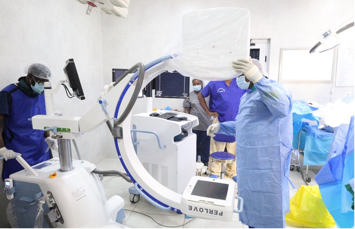
We are pleased to announce the introduction of a C-arm fluoroscopy machine to support orthopaedic operations in our theatre. This important investment strengthens our ability to provide image-guided surgical care and reflects our continuing commitment to modern technology, clinical excellence and patient safety.
The C-arm gives the surgical team access to real-time X-ray images during an operation. Instead of relying only on images taken before surgery, the surgeon can view bones, joints, surgical instruments and implants while the procedure is taking place. Fluoroscopy displays X-ray images on a monitor as they are produced, enabling clinicians to observe anatomy and the movement or placement of instruments in real time.
For patients undergoing selected orthopaedic procedures, this technology can provide valuable visual guidance at critical stages of an operation. It is not a replacement for the knowledge and judgment of the surgeon; rather, it is an advanced tool that helps the entire theatre team make well-informed decisions during surgery.
Understanding the C-Arm Machine
The equipment is known as a C-arm because its imaging components are connected by a curved arm shaped like the letter “C.” The design allows the machine to be positioned around the patient and adjusted to obtain images from different directions without repeatedly moving the patient. Mobile C-arm systems can be moved horizontally, vertically and around a pivot to create real-time images of bones and joints from the angles required by the surgical team (Meurer F, 2023).
On one side of the arm is an X-ray source, while the imaging detector is positioned on the opposite side. When an image is required, an X-ray beam passes through the part of the body being examined. The detector receives the information and sends the resulting image to a nearby monitor, where it can be viewed immediately by the surgeon and other authorized members of the clinical team.
Unlike a standard X-ray examination, which usually produces one or more still images, fluoroscopy can provide a sequence of images during a procedure. This gives the surgeon the ability to assess changes as they happen, including the alignment of a fracture or the position of an implant.
How the C-Arm Supports Orthopaedic Operations
Orthopaedic surgery frequently involves structures that cannot be seen directly from the outside of the body. During fracture treatment, for example, the surgeon may need to bring broken bone fragments into the correct alignment and then stabilize them using screws, plates, wires, rods or intramedullary nails.
Real-time fluoroscopic imaging helps the surgeon inspect the alignment of the bone and assess the location of instruments and implants before completing the operation. Orthopaedic fluoroscopy is used for procedural guidance and intraoperative decision-making in areas that include fracture fixation, spinal instrumentation, joint assessment, implant positioning and selected image-guided injections.
Depending on the patient’s condition and the procedure being performed, our C-arm may support operations involving:
- Reduction and fixation of fractures
- Placement of plates, screws, wires, pins and nails
- Assessment of bone and joint alignment
- Selected spinal procedures
- Guidance during some minimally invasive orthopaedic procedures
- Confirmation of implant positioning before the operation is completed
What the Technology Means for Patients
One of the most important advantages of the C-arm is that relevant images can be obtained inside the operating theatre. A radiographer or another appropriately trained and authorized professional operates the imaging equipment while working closely with the surgeon, theatre nurses and anesthesia team. Hospitals that provide theatre fluoroscopy use dynamic X-ray images to assist orthopaedic surgeons during procedures (Tischler et.al., 2015).
Immediate access to images allows the surgical team to assess the progress of an operation without waiting until the procedure has ended. When appropriate, the surgeon can use the images to refine bone alignment, adjust an instrument or reconsider an implant’s position before closing the surgical site.
For suitable procedures, image guidance can also support techniques that use smaller surgical access points because the surgeon can monitor internal structures on a screen rather than relying entirely on direct visual exposure. Fluoroscopy is widely used as guidance for interventional and minimally invasive procedures, although the size of an incision and the surgical approach remain dependent on the injury, the patient’s health and the operation required.
Strengthening Our Commitment to Modern Orthopaedic Care
The introduction of our C-arm machine represents an important step in the continued development of our orthopaedic and theatre services. It brings real-time imaging closer to the point of treatment and gives our surgical team another valuable resource for managing fractures, assessing alignment and guiding the placement of selected implants.
Most importantly, this investment is about people. It is about giving our clinicians access to appropriate technology, improving coordination in the operating theatre and providing patients with care informed by timely images and careful clinical judgment.
Every technology we introduce must serve a clear clinical purpose, be operated by competent professionals and contribute to a culture in which patient dignity, safety and quality of care come first. With the arrival of the C-arm, we are strengthening our capacity to deliver modern, image-guided orthopaedic surgery by combining skilled professionals, advanced technology and patient-centered care in one operating theatre.